.

.
marylin monroe
Showing posts with label paleo. Show all posts

The Potato Manifesto - Part 1/2: A (Re-)Evaluation of the Contemporary Discrimination of "the" Ordinary Potato

Image 1: The grunty regular (left) and the cute sweet potato (left), which one would you commit your health to? (img. menshealth.co.uk)
This is a blogpost, which eventually turned out to be the first part of a series, is a post with a history, a rather complex one, to be precise. It is rooted (not tubered ;-) in my amazement over the contemporary craving for sweet potatoes within the ever-growing neo-paleolithic community on the Internet and was sparked by the recent publication of a study on sweet potatoes, I stumbled upon on my daily tours of the most recent scientific literature. To make long story short, instead of immediately summarizing the data, drawing some graphs and commenting on the real-world implications of this study, I decided to use the holiday to descend into the archives and take a closer look at what science has to say on the bitter truth about the grunty regular and the starchy promises of cute sweet potato (cf. image 1 ;-)...

When wheat was devils excrement, would regular potatoes then be his horns?

According to the official obesity statistics of the European Union, British women are the fattest in Europe (Eurostat. 2011): 23.9% were classified as being obese (BMI > 30) in the year 2008 to 2009. This certainly raises the not altogether serious question, whether the British obesity problem (the obesity rate among the men was 22.1% and thusly topped only be the Maltese with 24.7%) is due to the fried fish or the fried (regular) potatoes in the unofficial British national dish, fish & chips.
Image 2: Obese British woman's rear view (img. BBC.co.uk)
A note to my dear American friends: Don't crow too soon, the obesity rate in the US tops the one in the UK by more than just a margin. According to a 2010 paper by Flegal et al. that was published in the Journal of the American Medical Association, the estimated obesity rate in the US amounts to 35.5% among the women and 32.2% among the men (Flegal. 2010). And a constantly increasing percantage (currently 4.7%) of the American population is already "extremely obese" and has a BMI > 40! Just to put that into perspective. A man or woman with a BMI beyond 40 and a height of 5 foot and 7 inches (170cm) would weigh at least (!) 254.85lbs (115.6kg) - this is already "Kig-Size Homer"-territory (cf. Intermittent Thoughts).
If we follow the current dietary paradigms and ignore the frying procedure, the answer to this question does not appear to be very difficult. I mean, when wheat was devil's excrement, then regular potatoes would be his horns or even nastier body parts, I do not want to mention here... and though a reasonable explanation for the widespread vilification of potatoes still escapes me, the contemporary nutritional paradigm within the health and nutrition blogosphere suggests that regular white potatoes have an awfully high glycemic index, will spike your blood sugar levels and have your neolithic body pump out tons of insulin - even if the rest of your diet is 99% paleo, as many of the listeners of Robb Wolf's podcast like to describe the way they are eating after having read his (widely read, yet controversial - esp. wrt to starches / carbs, fish oil and a few other topics) book ;-)
Figure 1: Names and characteristics of eight common potato cultivars in the British diet (left) and experimentally evaluated area under the glucose curve and glycemic indices (right; data based on Henry. 2005)
If we take a look at the data in figure 1, which shows both the incremental area under the glucose curve (AUC) and the GI of eight commercially available and commonly consumed British potato cultivars, it should however be quite obvious that the concept of the bad high-GI regular potato is about as misguided as the racist or religious prejudices some of our fellow human beings are still harboring against other members of the human race. With glycemic indices that range from 56 for the "waxy" Marfona to 94 for the "firm" Maris Peer, the "bad" regular potatoes cover the exact same GI range as their "healthy" sweet cousins (come back tomorrow for Part II of this series with more information on sweet potatoes).

Cultivar, processing, serving temperature and more have profound influences on the GI

The type  (=cultivar) of the (classic) potato, is yet neither the only, nor the most important determinant of the glycemic index of a potato meal. The processing method and, to my own surprise, even the food temperature have considerable influence on the glucose response to otherwise identical test meals, as well:
Figure 2: Incremental areas under the curve (AUC) and glycemic index values for 50 g available carbohydrate portions of white bread and seven potato meals tested in a cohort of 12 healthy subjects (data adapted from Fernandes. 2005)
On the left = "better than white bread"-side of figure 2, we have cold, boiled red potatoes (GI 56.2) and, surprise, frensh fries (GI 63.6) and roasted Californian white potatoes  (GI 72.3, but lower AUC than white bread). On the right = "worse than white bread"-side, instant mashed potatoes (GI 87.7) and the hot variety of the "low GI" red potatoes (GI 89.4) are competing for the red lantern.

With regard to the unexpected differences between cold and hot red potatoes, it is important to note that the results of a 2011 study by Kinnear et al. confirm that the latter is not an artifact of the Fernandes study. In their trial, the scientists from the University of Toronto found an average GI reduction of -37% (mean GI for the tested cultivars ~47), when the freshly boiled potatoes were refrigerated at 4°C for 24–28h before they were served to the 10 healthy study participants (Kinnear. 2011). As far as the reasons for this temperature-dependence of the glycemic index is concerned, Kinnear et al. speculate that it is an effect "due to starch retrogradation", which is a process that takes place in gelatinized starch, when the amylose and amylopectin chains realign themselves and thusly causes the liquid to gel. This is quite interesting, as it stands in line with the low GI of the Marfona potato (cf. figure 1), the texture of which is described as "waxy". The long-established relation of the phosphate content and the degree of starch gelatinization (and thusly digestibility and GI), on the other hand, could explain difference between crops and differences between identical crops grown on soil with different phosphate contents.
Figure 3: Areas under the glucose curves (AUC) in 32 healthy volunteers to 50g carbohydrates from mashed potatoes with or without 10g fructose administered 0, 30 or 60min before the meal. Measure by Accu Check finger-prick glucometer and YSI glucose oxidase analyzer (data adapted from Heacock. 2002)
Did you know that fructose, of all, is able to reduce the postprandial increase in serum glucose in nondiabetic adults (mean age: 26)? In their 2002 study, Patricia M. Heacock and her colleagues were able to show that pre-ingestion of 10g of fructose 60min and 30min before the ingestion of a 50g carbohydrate meal (from potatoes) reduced the area under the glucose curve (glucose AUC; cf. figure 3) by 25% and 27%, respectively (Heacock. 2002). The immediate co-administration of 10g of fructose with the potato meal (figure 3, 0 min), on the other, did not induce any statistically significant changes in the glucose AUC of the 13 male and 19 female study participants whose blood glucose levels were measured by finger-prick capillary blood (Accu Check) and glucose oxidase analyzers (YSI).
Furthermore, a 1999 study by Soh and Brand-Miller from the University of Sidney (Soh. 1999) shows that the real-world glucose responses of different individuals to differently processed and stored potato cultivars cover an even broader spectrum (especially on the low GI side) than the results of the previously cited studies suggested. The GI values, the Australian scientists calculated based on the glucose response to a 50g carbohydrate portion of eight different potato meals (three varieties, four cooking methods, two states of maturity) differed by as much as +/- 55pts, with canned new potatoes (GI 65) at the upper and boiled Desiree potatoes (GI 10) at the very lower end of the spectrum. And as if things were not complicated enough, already, Soh and Brand-Miller also introduce yet another variable into the equation - the size of the tuber, which showed a statistically significant correlation with the glucose response of the study 10 healthy participants (correlation between GI and tuber size: r=0.83, p < 0.05).

Black-and-white thinking and ineradicable prejudices

If we base our argumentation solely on the glycemic index, which is in fact the main argument that is brought forward against "regular" potatoes in the public debate, it is quite clear that the poor (regular) potato is another victim of the human propensity to black-and-white thinking and the public's stubborn adherence to convential nutritional wisdom. With reference to the "unjustified generalization" that "all potatoes have a high glycemic index", Anette E. Buyken and Anja Kroke, two researchers from the Research Institute of Child Nutrition in Dortmund, Germany, write in their letter to the editor of the British Journal of Nutrition (Buyken. 2005):
Figure 4: GI values (glucose reference) for potatoes by different cooking methods; the horizontal bars indicate the minimal and maximal glycemic index; the dotted vertical lines mark the conventionally accepted  "low GI" <55 (left) and "high GI" >70 (right) cut-off points (the figure was taken directly from Buyken. 2005)
[...] as with all GI data, the GI values of potatoes may depend on cooking method, processing, variety and the composition of the meal. This fact deserves attention since mashed potatoes, French fries, baked potatoes and potatoes cooked in a microwave are characterised by GI values mostly exceeding the upper limit for a high GI value of 70; whereas conventionally boiled potatoes appear to have a GI value on average below 70. The values of conventionally boiled potatoes do vary considerably though, so it may also be that some potato varieties have an inherently low GI what-ever the cooking method (Najjar. 2004; Fernandes. 2005). In this context, it should be considered that most currently available GI values are based on mature potato varieties (Ontario, Prince Edward Island, Desiree, Pontiac, Sebago). The starch of more mature potatoes is, however, easier to digest, presumably due to increased amylopectin branching and hence lower resistance to gelatinisation, which in turn results in a higher GI (Soh. 1999).
Buyken and Kroke support their argument by the means of an illustration of the broad range of the glycemic responses (and respective GI) of study participants to 46 different potato meals (cf. figure 4) and emphasize that there are significant differences in both the preferred potato cultivars, as well as the respective cooking / processing methods between European and US customers. While the former "prefer potato varieties characterised by a lower GI", the prevailing potato varieties in the US are mature and exhibit significantly higher glycemic indices. This trend toward higher glycemic indices in US potato meals is reinforced by the average American's preference to fry, bake, mash, roast or microwave his potatoes, so that it would  be "thoughtless" for any European, or American who selects less mature, low GI cultivars and refrains from frying, baking, mashing, roasting or microwaving his potatoes to follow the grossly over-generalized recommendation to eat less potatoes.

Ok, not all regular potatoes are made equal, but sweet potatoes are still king, right?

Against the background that my grand father who lived a 100% healthy, diabetes-free life into his late 90s, competed in track and fields and swam laps until about 6 months before he died, had regular potatoes with every dinner, I may be somewhat biased as far as the "bad potatoes" are concerned. This does yet not compromise the value of the rational arguments and scientific evidence against the unjust and, above all, over-generalized vilification of regular potatoes I have brought forward in this first installment of the Potato Manifesto. If you are interested in how the "holy" sweet potato which is currently hailed as the savior of the neo-paleolithic race compares, come back tomorrow for Part II of the Potato Manifesto ;-)

High Dietary Acid Load Doubles Risk of Type II Diabetes in Lean Individuals! Causative or Corollary? Plus: Are Grains, not Meats the Main Offenders in the Modern Diet?

If you go for green, you are usually on the save side of things... ah pHs ;-)
You will probably remember the association between higher acid levels (not even out of range), metabolic syndrome, lowered growth hormone release and a plethora of other ill-health effects I already hinted at in "How Could Bicarbonate Help You Lose Fat & Build Muscle" (read more). A new study from the Gustave Roussy Institute in France is now the first large scale study to provide relatively conclusive support for the hypothesis that there is a direct relationship between dietary acid load and the risk of cardiometabolic diseases and type II diabetes (Fagherazzi. 2013).

Credible evidence from large cohort

Fagherazzi et al. analyzed data from 66,486 women who were part of the E3N study (Etude Epidémiologique auprès des femmes de la Mutuelle Générale de l’Education Nationale), a French prospective cohort study of 98,995 female teachers, who were followed for incident diabetes over 14 years (the study started in 1999).
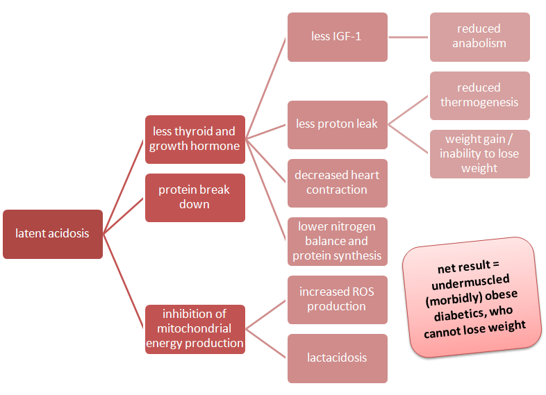
Latent Acidosis? Why do I care? As you can see in the image to the right, even latent acidosis can reduce thyroid hormone production, increase protein breakdown and inhibit mytochondrial function . The latter leads to increased ROS, lactate production & proton leak, while the former entails decreases in protein synthesis, IGF-1 & cardiac output. These changes have long thought to promote the development of the metabolic syndrome by increasing inflammation and reducing the basal metabolic rate (Berkemeyer. 2009).
During the 14-year follow-up period, a total of 1,372 cases of incident type 2 diabetes were validated. Fagherazzi et al.'s close analysis of the data revealed that there is a significant association between higher potential renal acid loads (PRALs) and the incidence of type 2 diabetes. Specifically, Fagharazzi et al. point out, ...
"[...] the highest PRAL quartile, reflecting a greater acid-forming potential, was associated with a significant increase in type 2 diabetes risk, compared with the first quartile (HR 1.56, 95% CI 1.29, 1.90)." (Fagherazzi. 2013)
A risk increase of 56%, alone, is a pretty impressive figure. What's even more impressive, though, is the fact that the association was significantly stronger among normal-weight women with a BMI <25 kg/m². For them the risk of developing type 2 diabetes almost doubles (+96%), when the dietary acid load is high. That's quite telling in view of the fact their baseline risk of developing T2D is low compared to those of the overweight study participants for whom the additional risk factor "dietary acid load" produced a significant (p = 0.03), but relatively low risk increase of only 28%.

The characteristics of the pro-diabetic diet

When we take a closer look at the actual data, there are dozens of statistically highly significant differences between the low and high pH quartiles (most of them with a p-value of p < 0.001). I initially tried to plot the differences, but that got way too chaotic, so I decided to make a list of items such as "higher energy intake (+15%)" indicating that the subjects with a high PRAL value (~the one's who were living on the more acidic side of the divide ;-) had a 15% higher energy intake than the subjects in the low PRAL quartile:
  • It may be coincidence, but in view of the anecdotal link between artificial sweeteners and heart-burn it's probably worth mentioning:  The most acidic study participants consumed 20% more artificially sweetened beverages (more about sweeteners)
    higher energy intake (+15%)
  • lower carbohydrate intake (-10%)
  • higher fat intake (+9%)
  • higher protein intake (+10%)
  • higher animal protein intake (+4%)*
  • lower fiber intake (-20%)
  • higher phosphorus intake (+16%)
  • lower potassium intake (-23%)
  • higher calcium intake (+10%)
  • lower magnesium intake (-22%)
  • higher sodium intake (+26%)
As a seasoned SuppVersity veteran, you will already have realized where this is heading: Higher energy intake, higher fat intake, hilariously low levels of potassium and magnesium, and salt intakes way beyond the 3g margin - all these are SAD (=standard American diet) hallmarks of the way the average Westerner (even in metropolitan France) eats. The 88% higher cheese intake, the 45% lower fruit + 25% lower veggie intake and 45% more of everyone's favorite "healthy" bread complete the picture that's emerging here: It's the Pizza Hut Diet ;-)

* The meat is not our only problem!

You will probably already have been wondering about the "*" and the fact that I highlighted higher animal protein intake (+4%)* in the previous list, right? Don't worry, I am not going to blame our problems on meat and suggest we all go vegan. The actual reason I highlighted the "bad" animal protein is a different one: the corresponding press release that came with the study (some of you may already have read it on Science Daily or other copy+paste 'science news portals'). It goes without saying that whoever wrote the short blurb used the study results for another sweeping blow at animal proteins by citing the following paragraph, and only but the following paragraph from the discussion of the results:
Suggested read: "Meat-Ology: The Link Between Red Meat, Cooking Techniques & Prostate Cancer" | more
"A diet rich in animal protein may favour net acid intake, while most fruits and vegetables form alkaline precursors that neutralise the acidity. Contrary to what is generally believed, most fruits such as peaches, apples, pears, bananas and even lemons and oranges actually reduce dietary acid load once the body has processed them. In our study, the fact that the association between both PRAL and NEAP scores and the risk of incident type 2 diabetes persisted after adjustment for dietary patterns, meat consumption and intake of fruit, vegetables, coffee and sweetened beverages suggests that dietary acids may play a specific role in promoting the development of type 2 diabetes, irrespective of the foods or drinks that provide the acidic or alkaline components."(Fagharazzi. 2013)
Obviously, it's going to be the first part of this paragraph, the one about the bad animal protein, that will get stuck in people's heads. The second part, the one that mentions rather casually that we are talking about total and not specific dietary acid loads, on the other hand, will go unnoticed. 

Figure 1: Food composition of paleo (top; estimation assumes a high meat intake) and modern US diet in % of total energy intake (Sebastian. 2013) - Don't forget: 1kg of lightly acidic foods are more acid forming than 100g of highly acidic foods!
Just as the fact that grains, which contribute an estimated 38% of the acid load yielded by the combined net acid-producing food groups in the contemporary diet (Sebastian. 2002), are just as, if not more problematic than the occasional steak of which the mainstream recommendations will tell you that you cannot have it more than once or twice a week, anyway.

'Paleo reasoning' to the rescue!?

In an effort to calculate the estimated net acid load of the 'ancestral', 'paleo' or 'whatever-you-want-to-call-it'-diet Sebastian et al. tested several scenarios, the worst of which a high protein, high fat version of the paleo diet (227g of protein, animal-fat content = 46%–63% of animal-food energy) still had a negative net endogenous acid production (NEAP = -7; Sebastian. 2013).

A low fat variety with a animal to plant food ratio of 35%:65% and an animal fat content of only 26% from animal-food energy and an even higher protein intake of 258g per day had a NEAP value of whopping -78. By "paleo standards" the average Westerner is thus consuming an extremely acidic diet, he is not genetically adapted to...  ;-)
Table 1: Acid-base value of common foods; positive values signify "acid forming", negative values "alkalizing" effects, the higher the figure the more pronounced the greater the impact of the given food is going to have (Cordain. 2012)
Paleo logic + scientific evidence = Win!  You all know that I don't buy the mainstream interpretation of the 'we are not adapted to...'-logic and the way it is employed by its followers in an almost religious way and against all scientific evidence.

What I do buy, however, are conclusions and recommendations that are based on both 'paleo logic' and scientific evidence - conclusions like the one Fagherazzi formulate in the last paragraph of their paper:
"[The] dietary acid load is directly associated with an increased risk of type 2 diabetes. From a public health perspective, dietary recommendations should not only incriminate specific food groups but also include recommendations on the overall quality of the diet, notably the need to maintain an adequate acid/base balance."
You want to know what foods Fagherazzi et al. may possibly be referring to? I already expected that. Just have a look at the table to the right, but don't forget: The study at hand does not prove causation. You could as well argue that it's the sum of dietary differences I listed as "characteristics of the pro-diabetic diet" which cause the diabetes. Anyways, the good news is: The solution, i.e. following a whole foods diet, is the same.

References:
  • Berkemeyer S. Acid-base balance and weight gain: are there crucial links via protein and organic acids in understanding obesity? Med Hypotheses. 2009 Sep;73(3):347-56.
  • Cordain L . AARP The Paleo Diet Revised: Lose Weight and Get Healthy by Eating the Foods You Were Designed to Eat. John Wiley & Sons, Apr 23, 2012
  • Sebastian A, Frassetto LA, Sellmeyer DE, Merriam RL, Morris RC Jr. Estimation of the net acid load of the diet of ancestral preagricultural Homo sapiens and their hominid ancestors. Am J Clin Nutr. 2002 Dec;76(6):1308-16.

Intermittent Thoughts On Intermittent Fasting - AMPK II/III: Leucine, HMB and a Glimpse on Other AMPK Modulators

Image 1: You pick a health, diet or diabetes supplement and I find the study that shows that in one way or another its effect is related to AMPK ;-)
I ended yesterday's installment of the Intermittent Thoughts on Intermittent Fasting Series on a pretty bold statement about the benefits of preworkout BCAA supplementation that would, at first sight, contradict common sense, or rather what common sense would dictate based on all you have read about the beneficial effects of BCAA supplementation on mTOR-related muscle protein synthesis (MPS) and the complementarity of mTOR and AMPK as regulators of anabolic (e.g. MPS, adipogensis ,etc.) and non-anabolic "scraping, rebuilding, recycling and repairing" processes. Since, after all, Bomb Jack, who posted a comment on last weeks installment of this series, is right: It would be logical that supplementation with BCAAs (he mentions HMB specifically) during the fast should result in dephosphorylation (~deactivation) of AMPK and thus negate its desirable effect on (metabolic) health.

And in fact, in the Wilson study I wrote about on Saturday the postprandial increase in AMPK phosphorylation, was blunted by the provision of carbohydrates, leucine or a combination of both (cf. yesterday's news) and you would assume that HMB supplementation would do the same, but the latter is - at least for chronic supplementation with low amounts (320mg/kg in rats ~ 52mg/kg in humans) of HMB - not the case (Pimentel. 2011), as the data I plotted in figure 1 clearly shows:
Figure 1: Effect one month of saline (control) or 80mg/day HMB on mTOR and AMPK phosphorylation and GLUT-4 expression in extensor digitorum longus (EDL) muscle of rats (Pimentel. 2011).
In the Pimentel study, there was, if anything, a non-significant increase in the AMPK and its purported downstream effect on GLUT-4 mediated glucose uptake  - both of which common sense would have told us to be compromised by HMB supplementation. While the lack of information on the "timing" or, more specifically, the interval between the last feeding and the intragastric administration (gavage) of 320 mg/kg body weight of HMB is a drawback in view of the significance of these results in an intermittent fasting context, rats usually eat at night and thus the administration of the 80mg of HMB (the rats weighed only 250g) "daily at the same time (during the light period)" will probably have coincided with a "fasting" period.

How can we explain that mTOR expression increased, while AMPK remained constant?

Are the different result a consequence of the metabolic magic of HMB? Well, before we analyze that in detail, there is another significant difference, we have to account for - in fact, a much more obvious one, which the amount of amino acids the rats were given in the Wilson and the Pimentel study, respectively (cf. figure 2).
Figure 2: Dosage, not type of supplement would be the most probable explanation for the different effects of leucine and HMB supplementation on AMPK phosphorylation in the Wilson vs. the Pimentol study.
I hope you did not already forget that, the main function of AMPK is to prevent that your cells run out of fuel or, to be precise, to avoid the ratio of "used" energy ADP and AMP (adenosine di- and monophospate) to ATP (adenosine triphospate) to continue to rise beyond a tolerable level. I further assume that you will be familiar with the fact that branched-chain amino acids bypass oxidation in the liver and thus become readily available energy sources for skeletal muscle (Renny. 2011). Now, if you put one and one together the answer seems pretty obvious: If the dosage of amino acids is sufficient (remember that those 270mg leucine are 4x more leucine than the the rats in the Wilson study got for "breakfast") to restore ATP levels to "appropriate" levels, the decrease in the ADP/ATP ratio will allow part of the AMP-activated protein kinase to be dephosphorylated.

According to our current understanding, BCAAs in general and leucine in particular trigger the ATP related decrease in AMPK and the complementary increase in mTOR by two distinct pathways, of which Tokunaga et al. write (Tokunaga. 2004)
[...]leucine stimulates p70α phosphorylation via mTOR pathway, in part, by serving both as a mitochondrial fuel through oxidative carboxylation and an allosteric activation of glutamate dehydrogenase. This hypothesis may support an idea in which leucine modulates mTOR function, in part by regulating mitochondrial function and AMPK.
In plain English: Leucine increases ATP when it is "burned" as fuel and it docks directly to the the non-active site of glutamate dihydrogenase enzyme and thusly increases the conversion of glutamate to alpha-ketoglutarate which in turn can be fed into the citric cycle to ultimately produce ATP.

Is it all about (cellular) energy ...

Figure 3: AMPK phosphorylation in Escherichia coli at different ADP/ATP ratios (data adapted from Xiao. 2011)
In April 2011 Xiao et al. published a study in Nature with some interesting quantitative data on the ADP/ATP ratio, on the one hand, the phosphorylation status of AMPK, on the other (Xiao. 2011). As my plot of the data in figure 3 shows, with increasing ATP levels (at constant ADP levels of 30µM) the phosphorylation of AMP-activated protein kinase in Escherichia coli BL21 cells declines by roughly -20% from 44% at a 30/0 ADP/ATP ratio to 22% at a 30/800 ADP/ATP ratio.

Yet, although these results would confirm the hypothesis that the main reason for the discrepancy is dose, or rather, energy related, and each and every nutrient that could potentially raise ATP levels, would eventually decrease AMPK, this still does not explain the increase in mTOR Pimentel et al. observed, despite (statistically non-significant) increases in AMPK.

... or is there a place for the "magic" of HMB?

As you probably know, beta-hydroxy-beta-methylbutyrat (HMB) is an oxidation product of leucine and / or its keto-acid alpha-ketoisocaproate (KIC) (Koevering. 1992). In 1998 Lembert et al. found that even KIC is not a direct substrate for ATP production, instead "KIC must transaminate with glutamate or glutamine to yield alpha-ketoglutarate and leucine" (Lembert. 1998). We may thus assume that similarly HMB cannot be used (directly) to restore cellular ATP pools. Moreover, HMB is thought to be the second (non-energetic) pathway by which leucine acts on protein synthesis / breakdown. According to a 2011 review of the literature by Zanchi et al. (Zanchi. 2011)
Nissen et al. (1996) suggested that HMB or some other metabolite (since there is no specific inhibitor to BCAT) is the main component responsible for the anti-catabolic effects of HMB because when adopting inhibitors of BCAA transamination, the only BCAA capable of anti-proteolytic effects is leucine, which undergoes a process capable of generating HMB (Slater and Jenkins 2000). Such effects were not observed when other BCAAs were tested (isoleucine and valine), suggesting that HMB or some metabolite may be the key element in promoting the [anticatabolic] effects.
When usually 5% of the dietary leucine is metabolized into HMB (Wilson. 2008), and these 5% are responsible for the non-ATP dependent effects on phosphorylation of mTOR, p70S6k, and 4E-BP1 of leucine (Eley. 2007), it is no wonder that chronic intake of 80mg of HMB did stimulate mTOR in the absence of increased ATP levels (which would obviously have led to a decrease in AMPK expression that was not present in the Pimentol study), while 270mg leucine, yielding only 13.5mg HMB, did not stimulate mTOR, but was (ab-)used as a substrate to increase cellular ATP levels, thusly reduced AMPK levels and increased protein anabolism - different pathways, similar results: an increase in net protein synthesis.
Figure 4: Simplified illustration of the two distinct pathways by which leucine can work its muscle protein synthetic (MPS) magic and a hint on the compensatory (/) / amplifying (+) effects of exercise.
There is however, a third major pathway to the metabolic effects that are brought about by common intermittent fasting programs and this third player makes things even more complicated (cf. figure 4) - it's exercise! You probably remember from yesterday's installment that
  1. during exercise in the fasted state temporarily AMPK increases and the energetically costly muscle protein synthesis (MPS) is reduced, while
  2. after exercise (regardless of whether it was performed fasted or not, cf. "Glycogen-Free Growth") muscle protein synthesis increases due to an exercise-induced stimulation of the mTOR protein synthetic cascade
Before we dig deeper into this modulatory effects of different modes of exercise in the next installment of the Intermittent Thoughts on Intermittent Fasting, however, I want to conclude today's thoughts with a preliminary list of supplements / medications that have been shown to modulate the phosphorylation state of 5' AMP-activated protein kinase.
Image 2: If you insist on trying HMB, don't be stupid and buy a capped products, the prices for bulk HMB powder have lately been crushed - a major European carrier, for example, sells 250g at <13€ atm; HMB is thus cheaper than BCAAs, which cost 16Euros in the small 250g pack - did you hear me say that even 13€ is too much, no - you must be mistaken ;-)
"Should you prefer HMB over leucine as a dietary supplement to promote lean mass gains and prevent muscle loss during the fast?" I assume this is a question many of you will now be pondering about. My answer to this question would be "NO!" Firstly, if you are no construction worker or pursue a similar physically demanding profession, the fear of losing muscle (which is different from "feeling flat", my bodybuilding friends ;-) during a ~16h fast is hilarious, which means that BCAA, Leucine or HMB supplementation, while you sitting fasted at your desk in the office is simply unwarranted. Secondly, when you are exercising the increased energy demand will negate / compensate the negative effect the increase in ATP has on AMPK activity. And thus, thirdly, a large bolus of leucine (or a complete BCAA or EAA product) taken pre-workout will not only ward off proteolysis (as HMB would do) it will also provide the necessary energy to train harder and thus help to increase the exercise induced stimulus on protein synthesis.

All that and the absence of conclusive scientific evidence that would demonstrate the superiority of HMB supplementation over the provision of adequately dosed BCAA or EAA mixtures (it stands to reason that you cannot compare 3g of HMB to 3g of BCAA) are arguments against the use of β-Hydroxy β-methylbutyric acid. If you wanted to try it, anyway (and have no problem swallowing a powder that tastes like poison), the prices for bulk-powders have gone through the floor, lately ;-)

How to modulate AMPK "artificially" -  supplements, medications, hormones and more

In view of the fact, that the discussion of the effects of leucine (BCAAs and HMB) alone took much longer than I had expected and this whole episode took a different turn than I would have expected, the following list is more a preliminary overview than a comprehensive explanation of the effects of various supplements, medications, hormones and hormone-like substances on the AMPK. The latter will follow, as promised, but for today, you will have to content yourselves with what I would like to call a sneak peak on the AMPK-mTOR modulation handbook of which I hope that it will be one of the outcomes of all the past and future work that is going into this series ;-)

AMPK promoters:
I still have two things to add to this list, firstly, this list is the result of a VERY cursory and 100% random search and is not even intended to be complete (at this time ;-). The intention (at least for in this installment) is to show you that an overwhelmingly large percentage of purported health supplements, diabetes and obesity treatments work via the AMPK pathway. And, secondly, I decided to limit the references to 1-3 per compound, even if in cases such as Metformin, ALA & Co the number of relevant studies is probably >500. Therefore you better consider the given references as evidence that I did not make up any associations between compound X and AMPK phosphorylation - and, if you want to know more before the release of the next installment, I suggest you go to PubMed and enter the respective keywords and do some digging on your own (your SuppVersity homework of the day - so to say ;-)

I hope you do not mind that I did not manage to tackle the effects of sleep and exercise in this installment, as I had originally intended. It is, after all, the central characteristic of this series that I sit down in front of the computer and start thinking at point "A", then I dig, here, get distracted there and follow up on "A1" to "A743", so that the output is by no means as structured and straight forward as my lectures and seminars or my SuppVersity blogposts on isolated topics... so, I can only hope that you enjoyed the turn this installment took (at best, because you learned something new) and in the unfortunate case that you did not enjoy what you have just read, you can at least look forward to the next episode of the Intermittent Thoughts on Intermittent Fasting Series ;-)

    Intermittent Thoughts On Intermittent Fasting - AMPK I/III: Zoning in on Its Effects on Body Composition

    Image 1: One reason why IF works is that it breaks the unnatural constant and convenient availability of high energy food and the subsequent suppression of AMPK phosphorylation (img courtesy of foxsearchlight)
    In the last installments of  this series we have begun to dig deeper into the signaling mechanisms that are / could be involved in the beneficial effects intermittent fasting is hailed for in the (unreal) world of the Internet blogosphere. We have identified AMPK and mTOR as the two players in the constructive and de-/re-constructive orchestrate of mammalian organisms and we have learned that their relationship - despite all its antagonistic aspects - is, after all, a complementary one. This means that, as you can observe it time and again in nature, health, vitality, yes even sustainable changes in body composition require balance!

    The delicate balance between mTOR and AMPK, this was another result of our considerations, is oftentimes broken in these days of nutritional abundance, where the rebuild and repair mechanisms of AMPK hardly get a chance to control the growth processes a constantly elevated mTOR pathway is triggering. The forced feeding-breaks on an intermittent fasting regime break this rampantly anabolic cycle. They let AMPK come into it's own and allow for...
    • ...broken DNA strands to be fixed, before their (re-)use results in cancerous growth (Habib. 2010),
    • ...cancer and defect cells to initiate apoptosis, i.e. to kill themselves (Chen. 2011)
    • ...life extension via reduction of dietary glucose to work (Schulz. 2007)
    • ...fat to be used as a substrate (Hardy. 2002), 
    • ...inhibiting adipogenesis = fat cell differntiation (Lee. 2011)
    • ...mitochondrial biogenesis to be initiated (Zong. 2002),
    • ...muscular GLUT4 activity and thus glucose uptake to be restored/increased (Holmes. 1999), 
    • ...gluconeogensis in the liver to be suppressed (Rutter. 2003), 
    • ...much much more healthy stuff ;-)

    AMPK may be non-anabolic, but that is not necessarily a bad thing!

    Image 2: Who would you prefer to be? Canto (left), on a life-extension (low calorie) diet with chronically elevated AMPK levels, or Owen (right), who enjoys his bananas to the fullest without even knowing about their profound effects on mTOR expression? (image taken from an article in the Irish Medical Times)
      
    On the other hand, we have also acknowledged the beneficial effects of mTOR on muscle growth, cell differentiation and many of the other processes that make life worth living and come to a hold, in the only physiological condition with chronically elevated AMPK levels, which is a "life" (if you want to call it such) extending low calorie diet, like the one poor Canto (image 2, left) has been on for a life, I personally would call miserable, when compared to that of Owen  (image 2, right) who obviously got his share of bananas.

    Moreover, studies on the effects of the AMPK inducing drug AICAR (cf. previous news on AICAR) suggest that the increase in mitochondrial oxidation may also lead to dangerously high levels of radical oxygen specimen (ROS) formation (Kim. 2006), of which you have learned only recently, that there is a fine line between the benefits of some vs. the deleterious effects of too much free radicals. Which brings up - yet again! - the issue of balance!

    Reversing perspectives: "Low energy" as the norm

    In the previous episode, we have also identified energy availability or, to be precise, the ratio of the high energy ATP (adenosine triphosphate) to the lower energy ADP (adenosine diphosphate; -7.3kcal/mol) and AMP (adenosine monophosphate; -10.9kcal), as a crucial determinant of AMPK activation. In one of the most recent reviews on the topic (Carlin. 2011), David Carlin and colleagues from the Imperial College in London state that
    [...] the finding that ADP, as well as AMP, protects AMPK against dephosphorylation influences the way we look at the physiological regulation of AMPK. To the best of our current understanding, the concentration of ADP in mammalian cells is much higher (10- to 100-fold)
    than that of free AMP, and so it is likely to be the main regulator of AMPK activity under normal energy-stress conditions. The extent of the tighter binding of ADP to AMPK, relative to MgATP, essentially offsets the higher physiological concentration of MgATP. An interesting possibility is that under most conditions AMPK is regulated by the ATP:ADP ratio through changes in Thr172 phosphorylation state. Under severe stress conditions, however, when the concentration of AMP might increase markedly, the additional allosteric activation mediated by AMP could act as a type of fail-safe device, ensuring that all AMPK substrates are maximally phosphorylated.
    In other words, under normal conditions ADP, i.e. the higher energy variety of the dephosphorylated ATP molecule, and not AMP is the main determinant of AMPK activity. And, and this is a pretty novel finding, both ADP and AMP do not actually activate 5' adenosine monophosphate-activated protein kinase (AMPK), but rather prevent it from being deactivated. While it may seem that this does not really matter, looking at things this way let's the "deactivation of AMPK by energy abundance" - a state we have accepted as a norm - suddenly look like the exception; and when we come to think of it, all the "diabesity"-related ailments an ever-increasing percentage of our society is experiencing can be tracked back to the reversal of norm (=AMPK phosphorylated = restore and repair using stored energy) and exception (=mTOR phosphorylated = build, grow, store) that is triggered by the persistent abundance of energy.

    Image 3: This is the way "fast food" is supposed to look like
    I think I do not have to tell you how to "restore" a normal state, which is characterized by recurrent episodes of increasing ADP levels: Exercise and fasting, or for the paleo crowd out there "hunting and gathering". I often make fun of the 1001 self-proclaimed paleo experts who try to explain everything based on largely non-verifiable assumptions about how our ancestors lived, but in this case, it is just plain obvious that "fast food", back in the day, was a rabbit that was too fast to be caught and not a hamburger, which, despite its greasy look, has about 2x more carbohydrates than fat (CalorieCount).

    What I am trying to say, here is that in human history, exercise or at least "movement" usually preceded nutrient availability. To facilitate that nature has equipped us with a compulsion to move most that is most prominent in anorexic patients, whose desire to "get going" is in part (another factor, these days, is obviously the hilarious calories-in-calories-out conception) mediated by the same mechanism that triggers the "food seeking behavior" in rats (Guisinger. 2003). With the neolithic age "food seeking" has become obsolete and with the advent of modern fast and convenient foods the movements we are making to avail us of the next (mostly sugary) snack or meal, whenever our body senses that the energy level is about to drop to normal (notice the change in perspective), takes us from the couch to the fridge and back... but I am digressing from the topic at hand, so let's get back to how intermittent fasting plays into that.

    Intermittent fasting = resisting the urge to go to the fridge

    Obviously your usual "walk" to the refrigerator is a definitive "no-no", when you are on in intermittent fast - or, to get back to the paleo metaphor, you are like Paleo Eve waiting for Paleo Adam to bring the "fast food" he is just chasing (the rabbit from image 3) home for you to roast it (I assume you will have read that doing the same with potatoes is not a good idea). While you are sitting there (on the couch or at the fire place, whatever you like better ;-) the majority of your 5' adenosine monophosphate-activated protein kinase will obviously stay in the same phosphorylated state it was, when you woke up this morning. This, in turn, brings up the question when / how the enzyme (AMPK) gets phosphorylated in the first place. A question you will probably be able to answer, if you read yesterday's news item on the effects of postprandial carbohydrate, leucine or carb + leu feeding on muscle protein synthesis.
    Figure 1: Postprandial changes in AMPK activity (relative to fasted state) 0min, 90min and 180min post ingestion of a 4g meal and following supplementation with water (control), carbohydrate (CHO), leucine (Leu), or leucine + carbohydrate 135min after the ingestion of the meal (data adapted from Wilson. 2011)
    If you take another look at the AMPK response to feeding and subsequent supplementation (135min post 4g of chow) you will notice that there is a decline in AMPK that corresponds to the peak in protein synthesis at 90min post ingestion of the meal. If you literally sit through (on the sofa or beyond the fire place without eating), AMPK will have reached it's former max again (and in the Wilson study peak even higher) another 90 minutes later. Thus the general recommendation to eat every 2-3 hours would leave little to no time for of normal / elevated AMPK levels during the day - this does certainly benefit muscle protein synthesis, as the latter - the results from the Wilson study confirm that - would be constantly elevated.

    In spite of the relatively rapid "restoration" of AMPK phosphorylation in response to "not eating", sitting on the sofa, watching TV and worrying about when you can finally break the fast is not what you would expect to give your physique the edge you are probably trying to achieve.
    Figure 2: Weight of fat pads in g/100g body weight in AICAR (intraperitoneal injections @ 0.7g/kg body weight) treated vs. control male Wistar rats after 4 and 8 weeks of treatment (data adapted from Gaidhu. 2011)
    On the other hand, Tipton et al. report that as you approach glycogen-depletion (your liver glycogen stores will be depleted within ~16h, cf. previous installments of the series), your basal AMPK activity will be elevated up to 2.5x (Tipton. 2006) - even if you are just sitting on the sofa! Moreover, chronic stimulation of AMPK via AICAR administration in a 2011 has been shown to have a exactly that beneficial modulatory effect on adipose tissue, that would be required for those type of changes, the health and fitness community generally labels as "recompositioning effects" (Ghaidu. 2011, cf. figure 2). Interestingly, the scientists did not observe any changes in lean mass (meaning you do not necessarily lose muscle when you deliberately increase AMPK), but - and this will remind you of the effects of high intensity interval training, you have read about, here at the SuppVersity earlier this week - chronically high levels of phosphorylated AMPK did increase mitochondrial density and energy expenditure (cf. figures 3-4).
    Figure 3: Inguinal mytochondrial density in AICAR
    (data adapted from Gaidhu. 2011)
    Figure 4: Energy expenditure in kcal/h
    (data adapted from Gaidhu. 2011)
    What would you say? Leaning out and setting the scene to stay lean by increasing the capacity to burn energy consequent to a >100% increase in mitochondrial density does not sound too bad, does it?

    Optimizing AMPK during the fast by exercise

    From previous installments of this series you know that the current "fitness-oriented interpretation of intermittent fasting", as I would like to call it, prescribes exercise as an obligatory 2nd element of a body recompositioning scheme à la leangains.com. It probably does not take a rocket scientists to gather that the energy depriving character of exercise will deplete cellular ATP levels, increase the APD:ATP ratio and thus prevent the dephosphorylation of AMPK. But if this is in fact the case (and it is, cf. "Exercise is perhaps the most powerful physiological activator of AMPK", Richter. 2009 ;-), the next question would be: How on earth can you still build muscle, if exercise activates AMPK and AMPK reduces protein synthesis? Well, the answer is simple: It's the seesaw principle!

    fast + exercise > AMPK up || rest + feed > AMPK down


    Figure 5: AMPK and protein synthetic response (relative to basal levels) to resistance exercise in 7 men and 4 women (data calculated based on Dreyer. 2006)
    In this context, the results of a 2006 study by Dreyer et al. (Dreyer. 2006) are quite exemplary. The scientists investigated the effects of resistance training in a fasted state (notice that AMPK is nothing only endless cardio sessions will promote) on AMPK phosphorylation and protein synthesis in 11 healthy young subjects. As the data in figure 5 goes to show, there was the expected increase in AMPK phosphorylation (+89% over baseline) during the exercise session, BUT despite constantly elevated AMPK levels, the protein synthesis, which had dropped to about 71% of baseline during exercise increased to +134% and +147% in the post-exercise period. This is initially counter-intuitive, because if AMPK were the sole deteriment of protein synthesis, the post-exercise rise in protein synthesis, Dreyer et al. observed should not have occurred. So how can we explain this "paradox"? The answer is pretty simple:

    Exercise is in itself a trigger for muscle protein synthesis (Drummond. 2009)!

    And if you remember the two posts on "glycogen-free muscle growth" related to the 2011 study by Camera et al. you will be aware that the phosphorylation of p70s6k and the subsequent increase in protein synthesis following resistance exercises does occur, even if you train in a glycogen-depleted state.

    Image 4: Duong at the beginning and 
    end of his 12-week intermittent fasting
    body transformation program;
    click here to read more
    Furthermore, the desirable (in terms of fat loss) increase in AMPK does not occur, if resistance training is performed in a fed state (Wittard. 2009). It should thus not be surprising that people like Duong (cf. image 4), in whose approach to intermittent fasting, training "on empty", i.e. at least without prior repletion of muscular glycogen stores by the consumption of great amounts of carbohydrates prior to his training sessions, was a staple, managed to achieve what everyone appears to be after these days, which is to lose fat and gain muscle (or at least maintain) muscle weight, at the same time! In that, the provision of supplemental BCAAs before resistance exercise (another of Duong's staples) could help prevent unwanted muscle breakdown, and increase the mTOR response without decreasing the exercise-induced increase in AMPK phosphorylation that is so beneficial in terms of metabolic health and, of course, fat loss.

    With that, I will leave you hungry for more until tomorrow, where due to a national holiday, here in Germany, I will have time to continue my elaborations on AMPK's role in intermittent fasting and beyond. So, I would suggest, you come back tomorrow if you want more information on how to modulate AMPK and its effects on your physique by exercise, sleep and supplementation to make your intermittent fast (or whatever other diet you are following) even more productive!

     update: Click here for part two...

    New Results From the "Test Tubers": Paleolithic Men Could Have Been Healthier Had They Microwaved Their Potatoes.

    Image 1: Potato roasting caveman-style - probably not the best way to "cook" your potatoes
    As a non-native-speaker, I must admit that the first time I heard someone talk about "tubers" was on Robb Wolf's famous podcast, back in the day, when I was "listener #6" (or seven ;-). Contrary to common (foreign) belief, not all Germans subsist on potatoes and sauerkraut and, what's more, even those who do, don't really care that a potato is a "tuber", i.e. a "Knolle" in German - I suppose the idea that it grows in the dirt is not too appealing to some, while the large majority probably just doesn't care as long as those "tubers" make a good addition to their Schweinebraten... yet, whatever the reasons may be, my first encounter with "tubers" has ingrained the link of these "underground structure[s] consisting of a solid thickened portion or outgrowth of a stem or rhizome, of a more or less rounded form, and bearing ‘eyes’ or buds from which new plants may arise" (OED.com), as the venerable Oxford English Dictionary would have it, to the paleo style of eating so deeply into my brain that I have been seeing cavemen with roasted sweet potatoes on their sticks in front of a fireplace in my mind's eye, ever since. Now, if that really was the way life went, back in the paleolithic days, our ancestors did probably miss about 32% of the antioxidant magic of the tuberous roots - at least this is what the results of a recent study on the effects of different cooking methods on polyphenols, pigments, and antioxidant activity in potato tubers from the San Luis Research Center at the Colorado State University  would suggest (Perla. 2011).

    In their experiment, Venu Perla, David G. Holm, and Sastry S. Jayanty
    • boiled - 1h in a sieved double-boiler,
    • microwaved + cooked - 10min in a commercial microwave oven at max. highest level +10min boiling, and
    • baked  - 1h at 204°C in a commercial pre-heated oven
    six months old stored potato tubers of 5 cultivars and 9 advanced selections of Colorado state (skin-color: 4x russet; 6x red; 1x white; 3x purple) and analyzed the samples for total phenolics, total flavonoids, total flavonols, and DPPH (2,2-Diphenyl-1-pikryl-hydrazyl) radical scavenging activity.
    Figure 1: Loss in total polyphenol content of 5 cultivars and 9 advanced selections of Colorado state potato tubers due to cooking, microwaving, and baking (data calculated based on Perla. 2011).
    As the data in figure 1 shows, all three preparation methods led to profound reductions of the potatos' total polyphenol counts. With Purple Majesty being most and Russet Nugget being least perceptible to the heat induced reduction in total polyphenol count. The cultivars CO97222-IR/R and CO97226-2R/R exhibited the highest total polyphenol counts (+77% and +137% above average in the raw state), with the former being particularly resistant to cooking (+119% above average) and the latter being particularly resistant to microwaving and baking (+154% and +169% above average, respectively).
    Figure 1: Loss in polyphenol, flavenoid and flavenol content of 5 cultivars and 9 advanced selections of Colorado state potato tubers due to cooking, microwaving, and baking (data calculated based on Perla. 2011).
    The superior resistance of these red-fleshed potato cultivars to cooking treatments is yet relative polyphenol-specific, due to their extraordinary high flavenoid and flavenol content in the raw state, CO97222-IR/R and CO97226-2R/R do yet retain a 241%, 155%, 199% and 243%, 331%, 395% higher flavenoid and 78%, 34%, 17% and 80%, 98%, 181% higher flavenol content  than the average potato (in the study) even after cooking, microwaving and baking, respectively.

    The scientists also observed that, contrary to the white and yellow fleshed tubers, where the major pigment was lutein, the "dominant pigments in the red and purple fleshed tubers were anthocyanins", the antioxidant activity of which has been implicated in the prevention (by some even the treatment) of obesity, cardiovascular disease, diabetes, hyperlipidemia and even cancer. Unfortunately, these pigments are just as susceptible to cooking, microwaving and baking as the polyphenols, flavenoids and flavenols, so that the total anti-oxidant activity (as measured by DPPH free radical scavenging assays) of the tubers that were tested in this study was reduced by -26%, -32% and -38% by boiling, microwaving and baking, respectively.
    Please note: While it is unlikely that you will die from eating a single raw potato. The solanine that can be found in all parts of the plant, including the leaves, fruit, and tubers, is a natural fungicide and pesticide the plant produces to protect itself from insects, the ingestion of which can potentially be fatal!
    Now, despite the fact that microwaving is only slightly less damaging that baking, I assume that putting the potatos directly into the fire, like the paloelithic men and women in my daydreams use to do it, probably is the worst way to prepare your tubers. Yet while even microwaving would have been a healthier option, the best and, from an evolutionary perspective, more natural solution would be to boil your potatoes.

    True or False? Glycine & Proline Supplements Ramp Up Collagen Synthesis & Improve Joint Health. Plus: The Tripeptide Advantage of Collagen Hydrolysates

    The "Paleo" cult has repopularized eating and preparing your own (Chicken) bone broth, but will this also help with bone and cartilage health?
    Although you're probably thinking of collagen as the stuff that's important for joint health, its implications in human health are more far-reaching than most of us believe.

    In fact, collagens are the most abundant group of organic macro-molecules in human and animal body. Because of their tensile strength, they perform numerous structural functions within the body - specifically in connective tissues which include among other tissue also organs as your heart, your intestines, your lungs and the parenchymal organs like the liver and the kidneys and even the fibrous matrix of skin and blood vessels.

    As I already said, collagens are yet by far best known as structural components of the protein matrix of the skeleton and its related structures, like bones, teeth, tendons, cartilage and ligament, which bring us back to the original question that bothered me after assuring Chris who emailed me asking about the necessity of taking glycine and proline supplements in the absence of any other protein (my answer was "that's bullocks"): Do glycine and problem supplements even help with collagen synthesis and joint health? Or is the supplement vendor next door the only person who benefits?
    You can find more True or False articles at the SuppVersity

    Pasta "Al Dente" = Anti-Diabetic

    Vinegar & Gums for Weight Loss

    Teflon Pans Will Kill You!

    Yohimbine Burns Stubborn Fat

    You Can Wash Pesticides Away

    High Volume Diet = Success
    We do have evidence (from rodent studies) that the ingestion of low molecular weight (=small peptides) collagen hydrolysates with intact glycyl-prolyl-hydroxyproline tripeptides that actually make it through the gut into the bloodstream and will increasee the organic substance content and decreased the water content of the left femur (Watanabe-Kamiyama. 2009). Previous studies had already shown hat the content of an orally administered gelatin hydrolysate will be incorporated into the cartilage tissue of rats (Oesser. 1999). Similar observations have been made by Iwai et al. for human volunteers and porcine gelatine hydrolysate, as well.
    "After the oral ingestion, the peptide form of Hyp significantly increased and reached a maximum level (20-60 nmol/mL of plasma) after 1-2 h and then decreased to half of the maximum level at 4 h after the ingestion. Major constituents of food-derived collagen peptides in human serum and plasma were identified as Pro-Hyp. In addition, small but significant amounts of Ala-Hyp, Ala-Hyp-Gly, ProHyp-Gly, Leu-Hyp, Ile-Hyp, and Phe-Hyp were contained." (Iawai. 2005)
    If we assume a similar physiological effect as it was observed by Watanabe-Kamiyama in rodents, the ingestion of (large) quantities of gelatine could thus very well, after it's hydrolysation in the gut, have similar effects on human cartilage tissue as the collagen hydrolysate that was used in the Watanabe-Kamiyama study.
    Table 1: Summary of Structure and Recovery of Food-Derived Collagen Peptide in Human Serum or Plasma after Oral Ingestion of Gelatin Hydrolysates (Iawai. 2005).
    With respect to the occurence of glycyl-polyl-hydroxproline tripeptides, of which the Watanabe-Kamiyama study suggests that they may be responsible for the beneficial effects on cartilage synthesis it should yet be said that it occurred in human plasma only after the ingestion of chicken, but not in porcine collagen in the Iawai study (see Table 1). If that's no coincidence, HARIBO, which is usually made with porcine gelatine is no "collagen builder", a real chicken soup, cooked with bone, on the other hand, could be.

    Given that your stomach is working properly a nice paleo bone broth (preferably from chicken bone) could thus produce similar results as a collagen hydrolysate of which a recent review in Current Medical Research and Opinion says that its ingestion stimulates a statistically significant increase in synthesis of extracellular matrix macromolecules by chondrocytes.
    There is more to collagen hydrolysates than joint health: In 2009 Saito et al. were able to show that fish collagen hydrolysates affect lipid absorption and metabolism in rats and may be useful in suppressing the transient increase of plasma triglycerides (Saito. 2009). Moreover, Spanish researchers showed that the daily dietary intake of hydrolyzed collagen seems to have a potential role in enhancing bone remodeling at key stages of growth and development in 60 children (9.42±1.31 years) who had been randomly assigned to either placebo or collagen (+ calcium) supplementation. In spite of these benefits, the ingestion of corresponding supplements is not necessary for people with healthy collagen metabolism who exercise regularly and eat clean.
    Figure 1: Physician rated (top) and subject-rated (bottom) improvement in joint pain walking (left) and standing (right) in the Clark study (Clark. 2008).
    The authors, researchers from the University of Illinois College of Medicine at Chicago and the University of Kiel in Germany add:
    "These findings suggest mechanisms that might help patients affected by joint disorders such as OA. Four open-label and three double-blind studies were identified and reviewed; although many of these studies did not provide key information – such as the statistical significance of the findings – they showed collagen hydrolysate to be safe and to provide improvement in some measures of pain and function in some men and women with OA or other arthritic conditions." (Bello. 2006)
    Subsequent studies such as Benito-Ruiz et al. (2009) or Clark et al. who evaluated data from 97 athletes from a varsity team or a club sport in Pennsylvania support Bello's conclusion (see Figure 1).

    Similar beneficial effects were also observed by  et al. in a more recent study with "normal" subjects with articular pain in response to 1,200mg/day of collagen hydrolysate (Bruyère. 2012). When we're looking into the effects of single amino acids, however, things look different. If they're ingested separately, glycine and proline are not going to form a tripeptide in the course of the digestive process. And while they may still serve as a raw material for the endogenous synthesis of such peptides the chance that they actively promote the synthesis of new collagen is slim.
    Biologically active tripeptides, not just glycine & proline is what you want!
    Bottom line: Collagen hydrolysates with intact tripeptides seem to have a beneficial effect on collagen synthesis. Classic broth and gelatine, both best made from chicken bones (absorption data on beef is not available), could have beneficial effects on collagen synthesis. In view of the chance that and rate at which the physiologically relevant  glycyl-prolyl-hydroxyproline tripeptides (see image to the right) are produced during the natural digestion process it does yet appear certain that you would have to garble down tons of it on a daily basis to actually trigger collagen synthesis and not just to do what individual amino acids could probably do as well: provide the necessary substrates without actually accelerating collagen synthesis.

    Chris' original question whether you'd have to take glycine and proline supplement on their own and in the absence of any other proteins and amino acids would thus actually be obsolete (you shouldn't take them at all), but I guess it may be worth mentioning that doing that, i.e. taking them on their own will only increase the "risk" of both being used by the liver as a substrate for glyconeogenesis (proline for example has the 3rd highest potential for gluconeogenesis 75% of the most glycogenic amino acid, i.e alanine; cf. Ross. 1967) - especially if you top "taking them on their own" with "taking them during a fast".
    References:
    • Bello, Alfonso E., and Steffen Oesser. "Collagen hydrolysate for the treatment of osteoarthritis and other joint disorders: a review of the literature." Current Medical Research and Opinion® 22.11 (2006): 2221-2232.
    • Benito-Ruiz, P., et al. "A randomized controlled trial on the efficacy and safety of a food ingredient, collagen hydrolysate, for improving joint comfort." International journal of food sciences and nutrition 60.S2 (2009): 99-113. 
    • Bruyère, Olivier, et al. "Effect of collagen hydrolysate in articular pain: a 6-month randomized, double-blind, placebo controlled study." Complementary therapies in medicine 20.3 (2012): 124-130.
    • Iwai, Koji, et al. "Identification of food-derived collagen peptides in human blood after oral ingestion of gelatin hydrolysates." Journal of agricultural and food chemistry 53.16 (2005): 6531-6536.
    • Oesser, Steffen, et al. "Oral administration of 14C labeled gelatin hydrolysate leads to an accumulation of radioactivity in cartilage of mice (C57/BL)." The Journal of nutrition 129.10 (1999): 1891-1895. 
    • Ross, B. D., R. Hems, and H. A. Krebs. "The rate of gluconeogenesis from various precursors in the perfused rat liver." Biochem. J 102 (1967): 942-951.
    • Saito, Masataka, et al. "Effect of collagen hydrolysates from salmon and trout skins on the lipid profile in rats." Journal of agricultural and food chemistry 57.21 (2009): 10477-10482.
    • Watanabe-Kamiyama, Mari, et al. "Absorption and effectiveness of orally administered low molecular weight collagen hydrolysate in rats." Journal of agricultural and food chemistry 58.2 (2009): 835-841.